

You are now on the AHDB Horticulture and Potatoes website.
Please click here to access the main AHDB website and other sectors.
Please click here to access the main AHDB website and other sectors.
The information was last updated in 2016.
Go back to the main page: narcissus leaf scorch, smoulder and white mould
Leaf scorch, caused by the fungus Peyronellaea curtisii (previously Stagonospora curtisii), is mainly a disease of Southwest England where it is favoured by the mild, wet weather conditions. It is most common on Tazetta types such as Grand Soleil d’Or and Innisidgen.
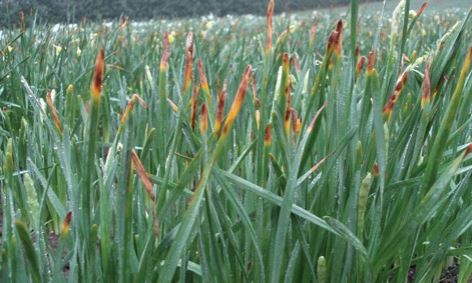
The name leaf scorch derives from the scorched or reddish-brown appearance of emerging leaf tips (see figures 1&2).
This initial symptom may easily be mistaken for frost or herbicide damage.
Later in the disease establishment, minute water-soaked spots with yellow centres develop. The centres become red-brown and necrotic as the lesions expand. These lesions remain small, rarely exceeding 2x5mm.
 ADAS
ADAS
Figure 1. Tip browning due to leaf scorch infection
 ADAS
ADAS
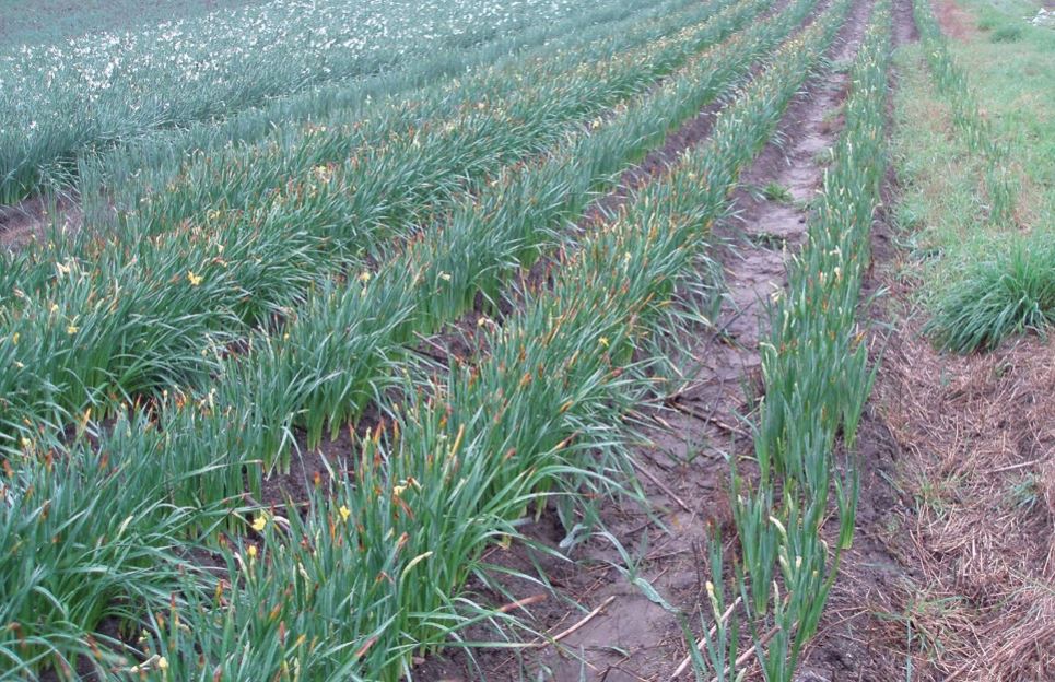
Figure 2. Widespread infection by leaf scorch in a susceptible variety
Although these brown elliptical spots remain relatively small, the leaf tissue above turns yellow and dies. Even in dried, withered leaves, the small brown elliptical spots can still be seen.
The transitional area between healthy and diseased leaf tissue is typically yellow.
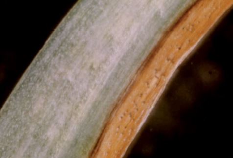
Fruiting bodies (pycnidia – see figure 3) develop in large numbers in the lesions, just visible to the naked eye as small brown swellings.
 ADAS
ADAS
Figure 3. Leaf scorch lesion with small, spherical pycnidia visible
The fungus also causes spots and blotches on flower stalks and flowers.
In susceptible varieties, leaf scorch can result in leaf dieback four to eight weeks before they should normally die; growers estimate that the resultant loss in bulb weight can be 30% or more.
It does not usually cause bulb decay.
The numerous spherical brown pycnidia (c.0.2mm diameter) within lesions serve to distinguish leaf scorch from smoulder and white mould - smoulder occasionally produces relatively large irregularly shaped black sclerotia within leaf lesions; whereas white mould produces numerous small (c.0.1mm diameter) black sclerotia.
Peyronellaea curtisii persists in or on dormant bulbs and this is considered the principal source of inoculum.
The fungus may be found between papery scales or between papery and fleshy scales.
Both the leaf tip scorch symptom and initial leaf spot infections (primary symptoms) take place as emerging leaves extend through the neck of the bulb.
Primary symptoms usually occur only on the outer leaves.
Pycnidia developing within primary symptoms contain a large number of spores, which are extruded when leaves are wet.
These spores are splashed or blown in wind-driven rain onto other leaves and flowers and onto neighbouring plants to cause fresh infections (secondary symptoms).
Peyronellaea curtisii can survive in old leaf debris for more than a year and this is likely to be an alternative source of initial inoculum.
Infested leaf debris could potentially initiate infection in following crops or increase disease spread in current crops.
Fungal mycelium of P. curtisii present in the bulb neck at planting infects emerging leaves.
Conditions that cause the pathogen to recommence growth are uncertain.
There is some evidence that the number of primaries in new plantings is associated with bulb storage conditions; storage at low temperature sometimes increases the problem, especially if combined with late planting.
The number of primaries does not appear to be related to the previous season’s foliar spread.
Wounding of emerging leaves is not necessary for infection to occur, although it is likely to increase disease development.
In experimental work on inoculated wounded leaves, water-soaked areas occurred within 24 hours, necrotic lesions within 48 hours and pycnidia within three days; on unwounded leaves the timescale was longer.
Wet conditions are required for rapid disease spread by spores.
Water on leaves causes mature pycnidia to swell and extrude spores; water splash results in spore spread from infected to healthy tissue; free water is necessary for the spores to germinate.
With moist conditions for 48 hours, spores result in small, pale spots after around four days and typical red brown elliptical spots after around 10 days.
Germinated spores produce infection hyphae, which penetrate the waxy cuticle; after this stage, the fungus is largely independent of external moisture.
Mycelial growth occurs between the cuticle and the epidermal cell wall.
Severe disease symptoms are most likely in wet weather and mild temperatures (15–20°C).
While leaf spots arising from bulb infection are located near the leaf tip, secondary leaf spots are formed over all parts of the leaf and can be especially numerous towards the base.
The mycelium in leaves grows down into the bulb neck as the leaves gradually die and very probably remains alive in the remnants of dead leaves during the bulb dormant period.
Topics:
Sectors:
Tags: